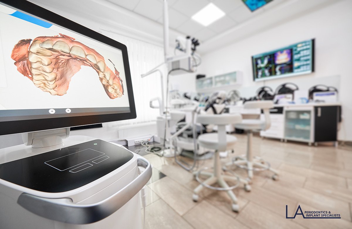
In-office 3D CBCT imaging gives our periodontists a complete digital view of your teeth, gums, and jawbone, so implant placement, bone grafting, and periodontal treatment are planned with far greater precision than 2D X-rays alone.
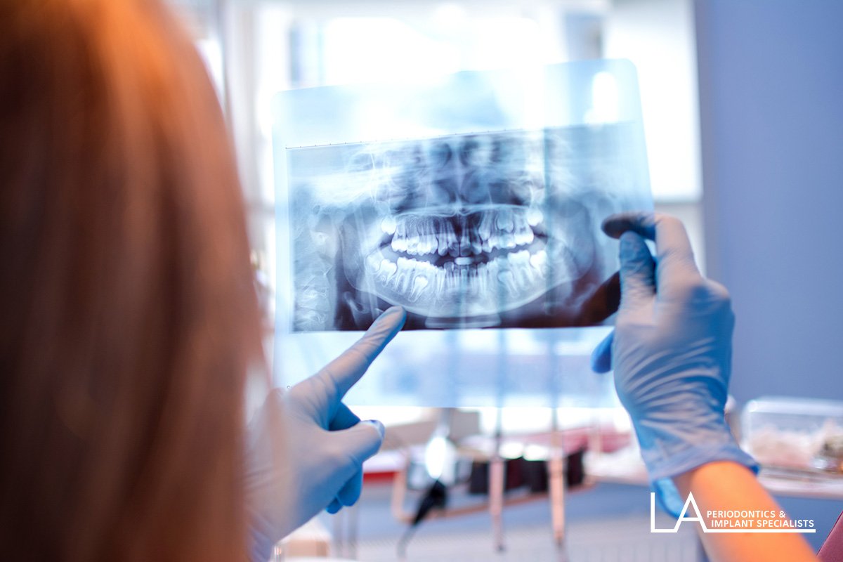
3D dental imaging, also called cone-beam computed tomography (CBCT), uses a rotating cone-shaped X-ray beam to capture your teeth, gums, nerves, and jawbone from every angle in a single scan. The result is a fully-rendered digital model, far more detailed than a traditional 2D X-ray, that our doctors use to plan treatment before a single incision is made.
Our practice uses in-office CBCT technology built for a lower effective radiation dose than many older machines, so you get high-resolution diagnostic detail with a fast, comfortable scan. Because everything is digital, results are ready to review the same visit, which means treatment planning for dental implants, bone grafting, and complex extractions moves forward without delay.
Your periodontist reviews your history and explains why 3D imaging is recommended for your specific treatment.
You sit or stand still while the scanner rotates around your head. Most scans take well under a minute and involve no contact or discomfort.
The 3D model is available immediately. Your doctor walks you through the images and points out exactly what they show.
Precise measurements of bone density, nerve position, and sinus location guide implant placement and surgical planning.
Every scan is reviewed by a board-certified periodontist, not just captured. Drs. Shahi, Young, and Yi interpret your imaging themselves.
Our Wilshire Blvd practice invests in in-office 3D imaging, so you are not referred out and left waiting on results.
Our imaging technology is designed for a lower effective radiation dose than many conventional machines, without sacrificing image detail.
Fully digital results mean your treatment plan can often begin the same day your scan is taken.
Dental implant planning. Precise bone measurements guide the size, angle, and position of every implant before surgery.
Bone graft evaluation. 3D imaging shows exactly how much bone is present, which determines the approach for a bone grafting procedure.
Diagnosing bone loss. Periodontal disease can quietly erode the bone that supports your teeth. 3D imaging reveals the extent of that loss.
Complex or full-arch cases. Cases such as All-on-4 implants and complicated extractions rely on 3D data to map nerves and sinus position safely.
Schedule a consultation to find out whether 3D dental imaging is recommended for your treatment plan. We welcome patients from throughout Los Angeles.
Yes. Dental X-rays, including 3D CBCT imaging, generate far less radiation than a chest X-ray, and the FDA considers dental imaging safe for almost all patients. We only order the scans that are necessary for your specific treatment plan.
The scan itself is quick, typically well under a minute. The scanner rotates around your head once to capture the full 3D data set, and your appointment includes time before and after to position you comfortably and review the images.
No. A CBCT scan is completely painless. You simply sit or stand still while the scanner moves around you. There is no contact, no injection, and no bite pressure involved.
A standard dental X-ray produces a flat, two-dimensional image. 3D CBCT imaging captures your teeth, nerves, and jawbone from every angle in one scan, giving your periodontist a complete model to measure bone density and plan procedures with much greater accuracy.
Coverage varies by plan and by the procedure the imaging supports. Our team verifies your benefits before your visit and will let you know what to expect. Call (310) 473-3800 and we can check your coverage in advance.
The 3D model shows exactly how much bone is available, where nerves and sinuses are located, and the ideal angle for each implant. This lets your periodontist plan implant placement digitally before surgery, which improves precision and reduces surprises during the procedure.
Not always. If a recent scan is available and still accurately reflects your current anatomy, your periodontist may be able to use it. A new scan is typically recommended when meaningful time has passed or when a procedure requires more current detail.
We'll confirm your visit within one business day.